Figure 1. [The normal human retina fundus]. - Webvision - NCBI
Por um escritor misterioso
Descrição
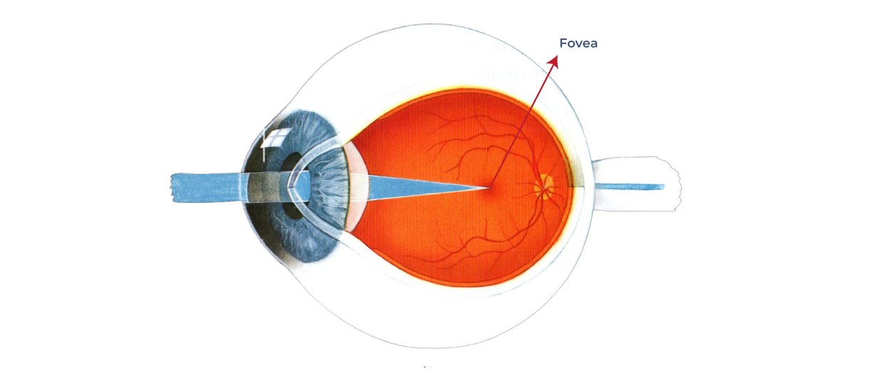
The normal human retina fundus photo shows the optic nerve (right), blood vessels and the position of the fovea (center).
![Figure 1. [The normal human retina fundus]. - Webvision - NCBI](http://webvision.instead-technologies.com/wp-content/uploads/2014/06/nervefibershuman1.jpg)
1.2 Simple Anatomy of the Retina. By Helga Kolb – Webvision
![Figure 1. [The normal human retina fundus]. - Webvision - NCBI](https://www.ncbi.nlm.nih.gov/books/NBK11533/bin/muller.gif)
Simple Anatomy of the Retina - Webvision - NCBI Bookshelf
![Figure 1. [The normal human retina fundus]. - Webvision - NCBI](http://eyerounds.org/atlas/LARGE/Normal-fundus-LRG.jpg)
Atlas Entry - Situs Inversus of the Retinal Vessels
![Figure 1. [The normal human retina fundus]. - Webvision - NCBI](https://www.ncbi.nlm.nih.gov/books/NBK11556/bin/factsf6.gif)
Facts and Figures Concerning the Human Retina - Webvision - NCBI Bookshelf
![Figure 1. [The normal human retina fundus]. - Webvision - NCBI](https://journals.physiology.org/cms/10.1152/physrev.00035.2019/asset/images/medium/z9j004202952r001.png)
Emerging Approaches for Restoration of Hearing and Vision
![Figure 1. [The normal human retina fundus]. - Webvision - NCBI](https://www.researchgate.net/publication/242466981/figure/fig1/AS:298496365219842@1448178487460/A-Normal-fundus-of-OD-B-Fundus-of-OS-showing-foveal-retinal-pigment-epithelial.png)
A) Normal fundus of OD; (B) Fundus of OS showing foveal retinal
![Figure 1. [The normal human retina fundus]. - Webvision - NCBI](https://eophtha.com/images/uploads/159747450519834383625f3786c924269.jpg)
Anatomy of Retina
![Figure 1. [The normal human retina fundus]. - Webvision - NCBI](https://media.springernature.com/lw685/springer-static/image/art%3A10.1038%2Fs41467-019-12917-9/MediaObjects/41467_2019_12917_Fig4_HTML.png)
Single-nuclei RNA-seq on human retinal tissue provides improved transcriptome profiling
![Figure 1. [The normal human retina fundus]. - Webvision - NCBI](https://www.mdpi.com/cells/cells-12-01987/article_deploy/html/images/cells-12-01987-g001.png)
Cells, Free Full-Text
de
por adulto (o preço varia de acordo com o tamanho do grupo)