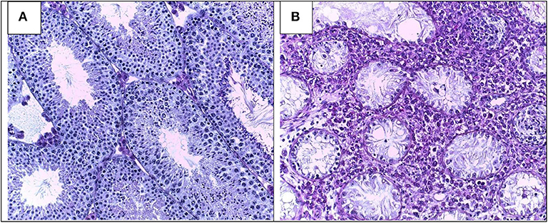
Morphology of Leydig cells in the testes after in vivo MCP-1 treatment.
Por um escritor misterioso
Descrição
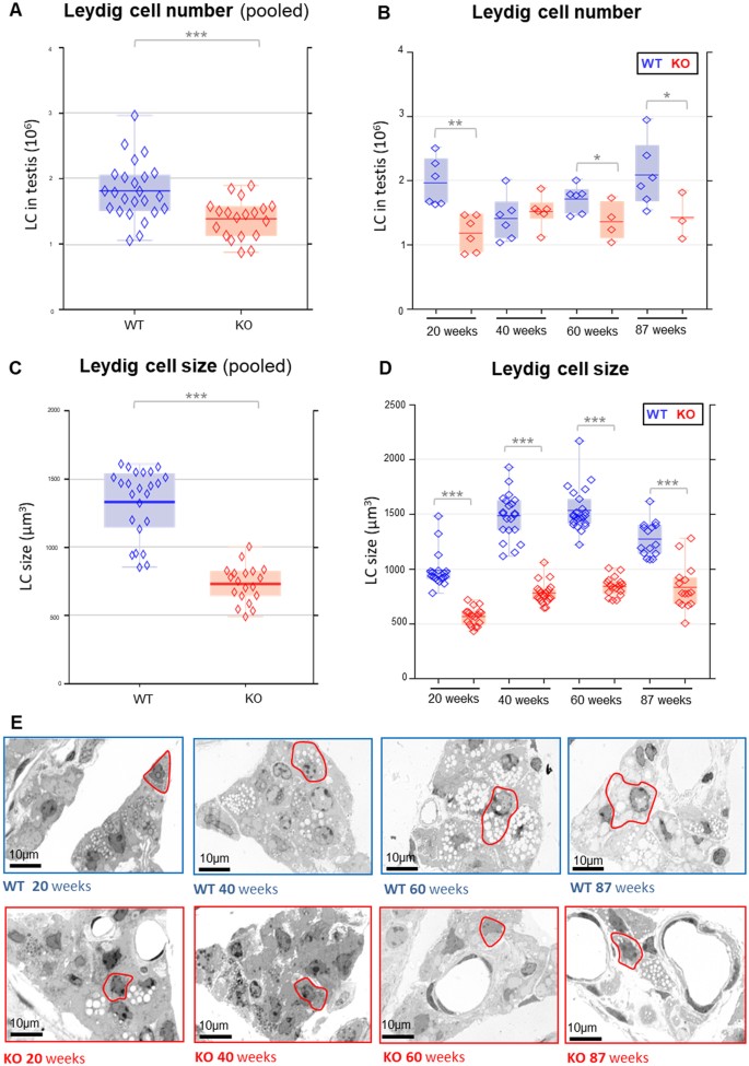
Low testosterone in ApoE/LDL receptor double-knockout mice is associated with rarefied testicular capillaries together with fewer and smaller Leydig cells

Morphology of Leydig cells in the testes after in vivo PTHrP
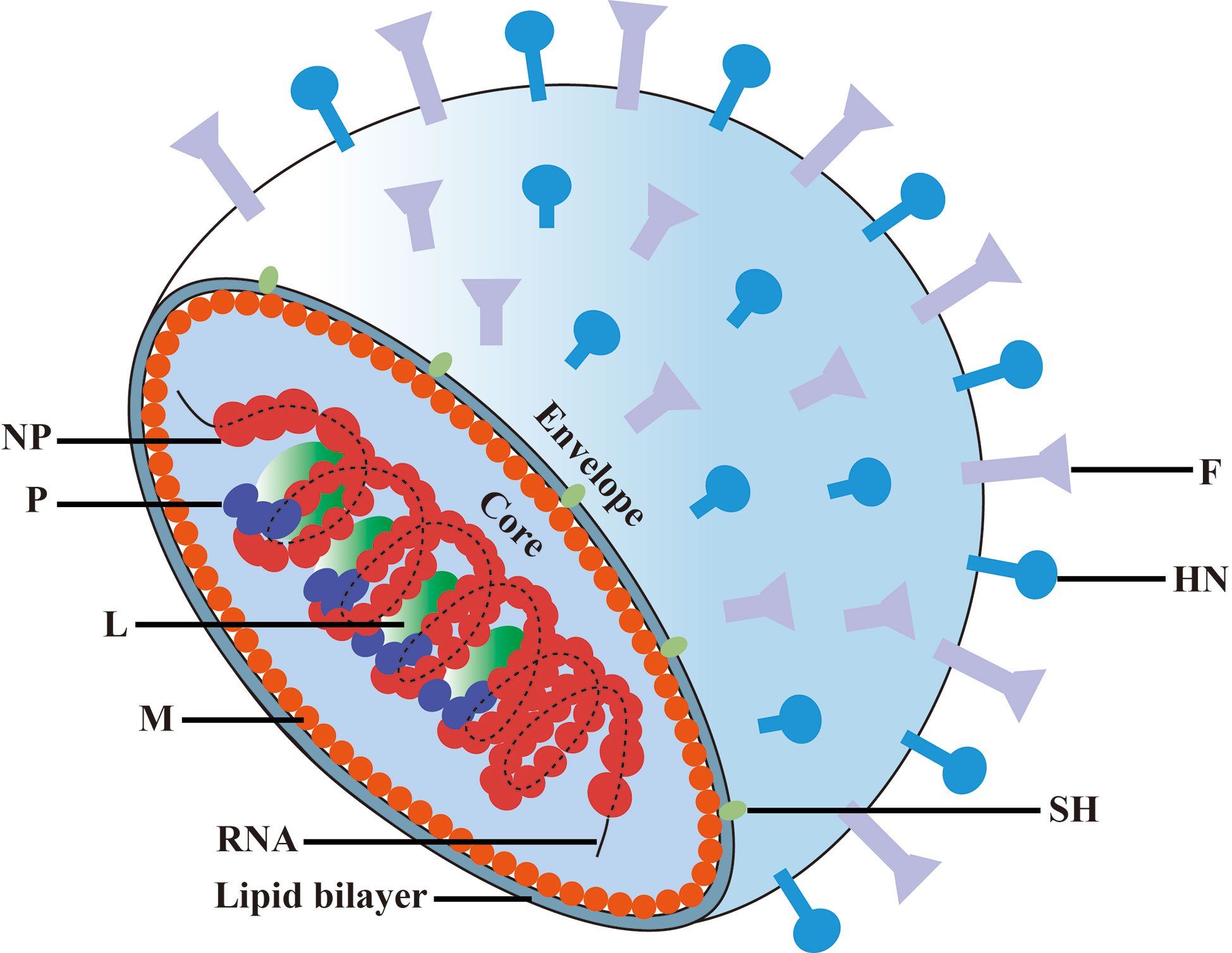
Frontiers Mumps Orchitis: Clinical Aspects and Mechanisms
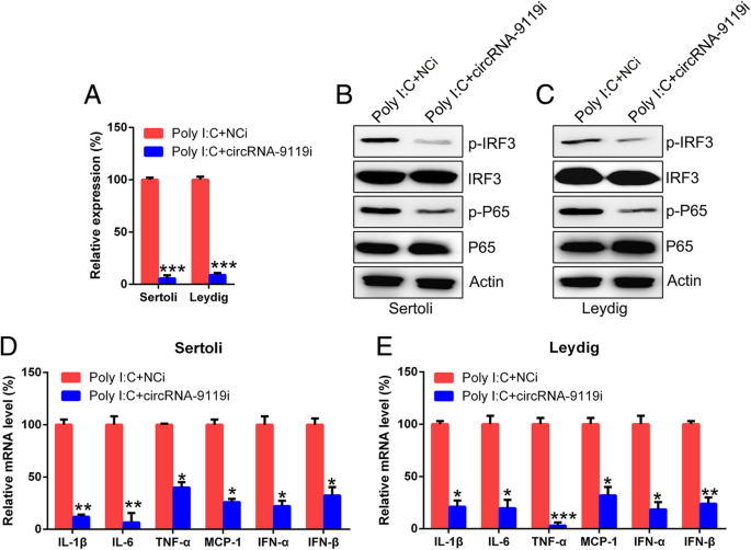
CircRNA-9119 suppresses poly I:C induced inflammation in Leydig and Sertoli cells via TLR3 and RIG-I signal pathways, Molecular Medicine
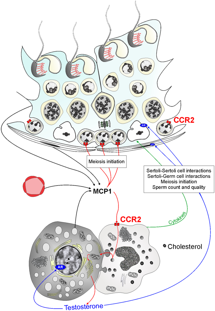
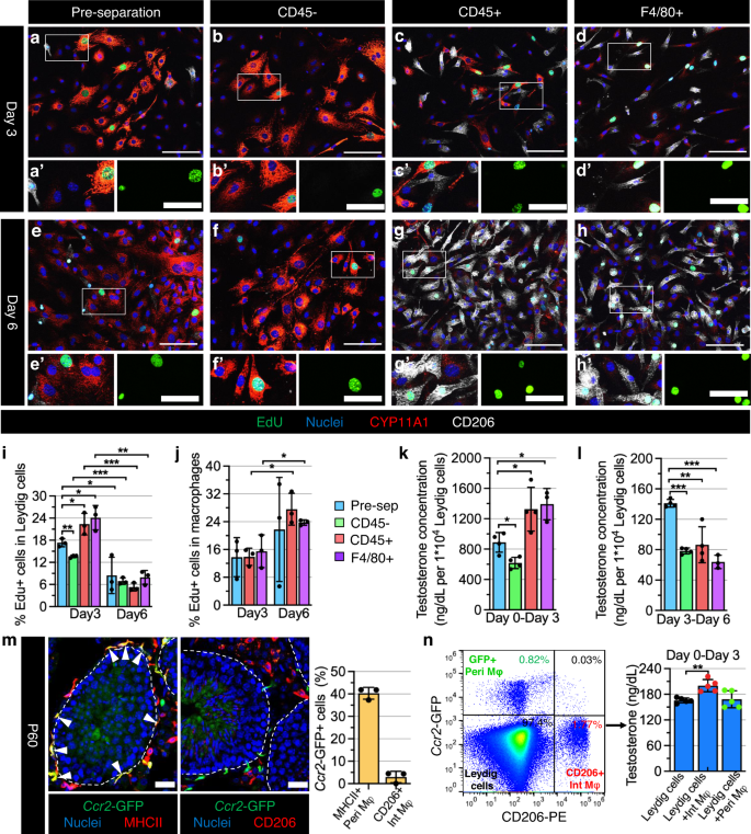
Activation of C–C motif chemokine receptor 2 modulates testicular macrophages number, steroidogenesis, and spermatogenesis progression
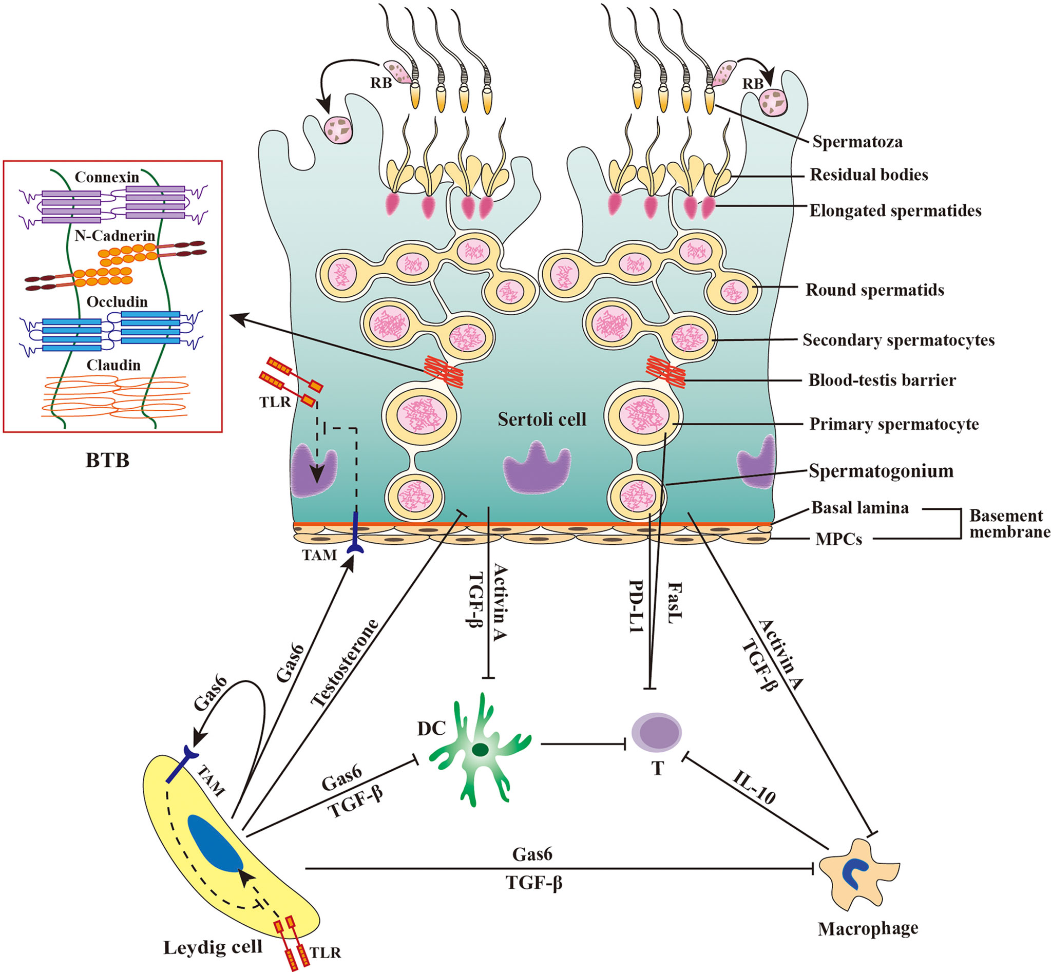
Frontiers Pathomechanisms of Autoimmune Based Testicular Inflammation
Testicular macrophages are recruited during a narrow fetal time window and promote organ-specific developmental functions
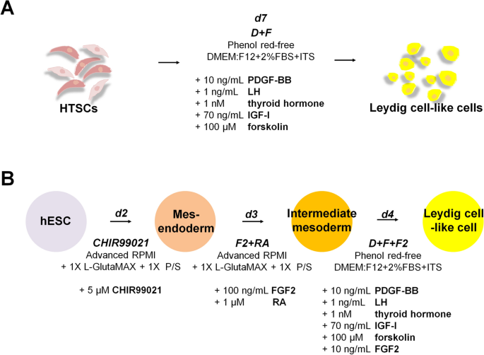
Rapid Differentiation of Human Embryonic Stem Cells into Testosterone-Producing Leydig Cell-Like Cells In vitro
Frontiers Viral tropism for the testis and sexual transmission

Stem Leydig cells: Current research and future prospects of regenerative medicine of male reproductive health - ScienceDirect

Testicular torsion in vivo models: Mechanisms and treatments - Minas - 2023 - Andrology - Wiley Online Library
IJMS, Free Full-Text

Impact of Toxoplasma gondii infection on TM3 Leydig cells: Alterations in testosterone and cytokines levels - ScienceDirect
de
por adulto (o preço varia de acordo com o tamanho do grupo)