Topographic sketch. Coronal view of bilateral DRT (orange). Patient
Por um escritor misterioso
Descrição

PDF) Three-dimensional LASIK flap thickness variability: Topographic central, paracentral and peripheral assessment, in flaps created by a mechanical microkeratome (M2) and two different femtosecond lasers (FS60 and FS200)

JCM April-2 2023 - Browse Articles

DTI for brain targeting: Diffusion weighted imaging fiber tractography—Assisted deep brain stimulation - ScienceDirect

e‐Poster - 2023 - Clinical Oral Implants Research - Wiley Online Library
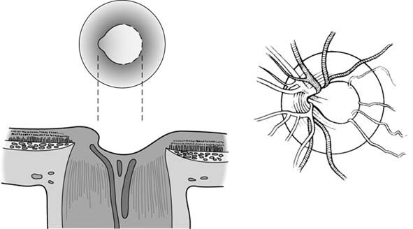
AccessLange: General Ophthalmology ; Chapter 2: Ophthalmologic Examination, Page 1

Single neurons and networks in the claustrum integrate input from widespread cortical sources - Abstract - Europe PMC

An atlas of white matter anatomy, its variability, and reproducibility based on Constrained Spherical Deconvolution of diffusion MRI

Promises of Functionally Graded Material in Bone Regeneration: Current Trends, Properties, and Challenges

Crossing nerve transfer drives sensory input–dependent plasticity for motor recovery after brain injury

Neuro-Ophthalmology & Neuro-Otology, Media

Subthalamotomy for Parkinson's disease: clinical outcome and topography of lesions
de
por adulto (o preço varia de acordo com o tamanho do grupo)